In the intricate network of the human circulatory system, countless veins play a vital role. Among these, the median cubital vein stands out for its accessibility and clinical significance, particularly in the field of phlebotomy. This guide, designed specifically for Phlebotomy Now School students, delves into the anatomy, structure, location, and clinical applications of this crucial vein.
| Key Takeaways |
| The median cubital vein is the most prominent superficial vein in the body, ideally suited for blood draws and IV access. |
| Its large size, stability, and minimal surrounding nerves make it a preferred choice for phlebotomists. |
| Understanding anatomical variations of the median cubital vein is crucial for successful phlebotomy procedures. |
| Proper tourniquet application and aseptic technique are essential for safe blood draws and IV cannulation. |
Understanding the Body’s Circulatory System: Arteries, Veins, and Capillaries
Before zooming in on the median cubital vein, let’s establish a strong foundation in the body’s circulatory system. This system comprises arteries, veins, and capillaries, working together to deliver oxygen-rich blood to tissues and transport deoxygenated blood back to the heart and lungs for replenishment [2]. Arteries, like highways, carry oxygenated blood away from the heart. Veins, on the other hand, function as return vessels, carrying deoxygenated blood back to the heart. Capillaries act as the exchange points, allowing oxygen and nutrients to move from the blood to tissues and waste products to move from tissues back into the blood.
Why the Median Cubital Vein is Preferred for Venipuncture
The median cubital vein, also known as the median basilic vein, reigns supreme as the most prominent superficial vein in the body. It’s readily visible on most individuals at the inner fold of the elbow, nestled within the cubital fossa (the triangular depression of the elbow joint) [3]. This superficial positioning, meaning it lies close to the skin’s surface, makes it the preferred choice for phlebotomists performing venipuncture (blood draws) and intravenous (IV) cannulation.
Anatomy of the Median Cubital Vein: From Lumen to Adventitia
Similar to most veins, the median cubital vein boasts a multi-layered structure, each layer contributing to its function:
- Lumen: The inner opening of the vein, typically ranging from 2.3 to 4.9 millimeters (mm) in diameter [4].
- Valves: One-way valves that prevent blood from flowing backward and ensure unidirectional flow towards the heart.
- Endothelium: A single layer of cells lining the inner surface of the vein, playing a crucial role in blood flow regulation and preventing clot formation.
- Basement Membrane: A thin layer separating the endothelium from the surrounding smooth muscle.
- Smooth Muscle: This muscular layer contracts and relaxes to regulate blood flow within the vein.
- Adventitia: The outermost connective tissue layer, providing structural support and anchoring the vein to surrounding tissues [3].
How to Locate the Median Cubital Vein in the Arm
The median cubital vein bridges the gap between the two major superficial veins of the upper arm:
- Basilic Vein: This vein originates on the outer hand near the pinky finger and travels upwards to the armpit (axilla) [5].
- Cephalic Vein: Beginning on the inner hand near the thumb, it courses upwards to the outer top of the shoulder [5].
Both the basilic and cephalic veins ultimately drain into the axillary vein. The median cubital vein, typically positioned diagonally, connects these two veins at the inner elbow, lying approximately 2–3 mm below the skin’s surface [4].
Understanding Anatomical Variations of the Median Cubital Vein
The median cubital vein exhibits some fascinating anatomical variations. While the standard configuration involves an upward diagonal course connecting the basilic and cephalic veins, research suggests that only about half of adults possess this pattern [6]. In some individuals, the basilic or cephalic vein might deviate from the usual path, potentially even eliminating the connecting median cephalic vein altogether.
The absence of a median cephalic vein, though uncommon, can occur in both men and women. Additionally, rare cases of individuals with two median cubital veins have been documented. However, further research is needed to determine the prevalence of this variation.
Role of the Median Cubital Vein in the Circulatory System
The primary function of the median cubital vein, like all veins in the arm, is to return deoxygenated blood to the heart and lungs for oxygen replenishment. The basilic vein carries the majority of the blood volume back to the lungs, with the median cubital vein facilitating efficient drainage between the basilic and cephalic veins.
Why the Median Cubital Vein is Essential for Phlebotomists
The median cubital vein, while not essential for survival, significantly aids venous return from the arms. However, its true significance lies in its application within phlebotomy. Due to its prominent visibility and superficial location, the median cubital vein serves as the preferred site for venipuncture (blood draws) and intravenous (IV) cannulation for several reasons:
- Size and Stability: The median cubital vein boasts a larger diameter compared to other peripheral veins, making it easier to locate and access with a needle. Additionally, its positioning minimizes movement or rolling of the vein during needle insertion, reducing the risk of unsuccessful attempts and patient discomfort [4].
- Reduced Pain: The area surrounding the median cubital vein contains fewer nerve endings, minimizing pain associated with venipuncture. The patient will thus have a more comfortable experience.
- Nerve Protection: The positioning of the median cubital vein, overlying arterial vessels, offers a layer of protection for these vital structures during venipuncture when the arm is extended and the inner elbow skin is taut [4].
Tips for Successful Venipuncture Using the Median Cubital Vein
While the median cubital vein presents numerous advantages for phlebotomists, certain factors require consideration to ensure a successful and safe procedure:
- Patient Anatomy: As discussed earlier, anatomical variations in the median cubital vein exist. Phlebotomists must be adept at palpating (feeling) for the vein’s location and course to avoid complications. In cases where the median cubital vein is absent or unsuitable, alternative veins in the arm, such as the cephalic or basilic vein, can be used.
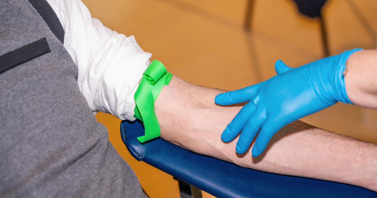
- Tourniquet Application: Applying a tourniquet properly is crucial for successful venipuncture. The tourniquet should be placed upstream (closer to the heart) from the chosen venipuncture site and tightened to a degree that restricts venous flow but not arterial flow. Once blood flow is visualized in the vein below the tourniquet, it can be loosened slightly to facilitate needle insertion.
- Aseptic Technique: Maintaining sterile technique throughout the venipuncture procedure is paramount to prevent infection. This involves proper hand hygiene, disinfection of the venipuncture site, and using sterile equipment.
Common Conditions That Can Affect the Median Cubital Vein
The median cubital vein, while a workhorse for phlebotomists, is not invincible. Certain conditions can threaten its functionality and complicate blood draws. Let’s delve into these conditions and explore how to maintain a healthy median cubital vein.
Recognizing and Managing Common Vein Conditions
- Phlebitis: Inflammation of the vein wall, often caused by irritation from intravenous (IV) catheters or infections. Symptoms include redness, pain, and tenderness along the vein.
- Thrombophlebitis: This condition combines phlebitis with blood clot formation (thrombus) within the inflamed vein. It can cause swelling, warmth, and redness in the affected area, and potentially limit blood flow.
- Median Cubital Vein Thrombosis: A specific case of thrombophlebitis isolated to the median cubital vein. It can significantly hinder blood draw attempts in this location.
Early detection is crucial for effectively managing these conditions. If you experience any redness, pain, or swelling near your elbow after a blood draw or IV insertion, consult your doctor immediately.
How to Keep Your Median Cubital Vein Healthy
- Minimize needle sticks: Repeated venipuncture attempts can irritate the vein. If phlebotomists struggle to access the median cubital vein, alternative veins in the arm can be used.
- Hydration is key: Adequate fluid intake plumps veins, making them easier to locate and access during venipuncture. Try to drink eight glasses of water every day.
- Warm compresses: Applying a warm compress to the inner elbow before a blood draw can help dilate the vein, facilitating easier access.
- Maintain healthy habits: Exercise, a balanced diet, and managing stress all contribute to good overall vascular health, including the median cubital vein.
Other Venipuncture Sites When the Median Cubital Vein is Unavailable
If the median cubital vein is inaccessible due to scarring, thrombosis, or other reasons, phlebotomists can utilize alternative access points:
- Peripheral intravenous access: This involves using other veins in the arm or hand, such as the cephalic vein or basilic vein.
- Central venous access: In rare cases, for critical situations or long-term IV therapy, larger veins in the chest (central venous access) might be used.
Other Uses of the Median Cubital Vein in Medical Procedures
Beyond blood draws, the median cubital vein can be used for:
- Blood cultures: Extracting blood samples for identifying bacterial or fungal infections.
- Administration of medications or fluids: Delivering medications or fluids directly into the bloodstream through an IV line.
Tips for Patients to Ensure a Successful Blood Draw
- Holding still: Minimize movement during venipuncture to reduce the risk of vein puncture and discomfort.
- Understanding the procedure: Knowing what to expect during venipuncture can alleviate anxiety and promote a smoother experience.
- Reducing the risk of fainting: If you’re prone to fainting during blood draws, inform the phlebotomist beforehand. Techniques like lying down or tensing leg muscles can help.
By understanding potential issues and following these tips, you can ensure the continued health of your median cubital vein and contribute to a smooth and successful blood draw experience.
FAQs: Common Questions About Blood Draws
Can blood be drawn from other veins besides the median cubital vein?
Yes, if the median cubital vein is absent or inaccessible, other veins in the arm, such as the cephalic or basilic vein, can be used for blood draws.
What should I do if I experience pain during venipuncture?
Inform the phlebotomist immediately. They can adjust their technique or choose a different vein to minimize discomfort.
Are there any risks associated with venipuncture?
While generally safe, venipuncture carries minimal risks like bruising, bleeding, or infection. Proper technique and aseptic precautions significantly reduce these risks.
Conclusion: A Phlebotomist’s Ally
The median cubital vein stands out as an invaluable asset for phlebotomists. Its accessibility, size, and stability make it the preferred choice for venipuncture and IV cannulation. By understanding its anatomy, variations, and proper access techniques, phlebotomists can ensure smooth, efficient, and comfortable procedures for their patients.
Phlebotomy Now School: Equipping You for Phlebotomy Success
Phlebotomy Now School empowers aspiring phlebotomists with the knowledge and skills necessary to excel in this rewarding field.
Our comprehensive curriculum delves into venipuncture techniques, proper needle selection, and best practices for ensuring patient comfort and safety.
With a focus on the median cubital vein and other relevant anatomy, Phlebotomy Now School equips you to confidently navigate the world of phlebotomy.